cvi42’s Cardiac MR offering is a one stop shop for all your clinical CMR needs. Quantify cardiac function, flow and assess tissue abnormalities faster than ever before with AI-based contouring.
Function
Function
Increase scanning throughput for myocardial function with quick ejection fraction, stroke volume and mass calculations. Reduce manual workload with accurate and reproducible volumetric assessment for all cardiac chambers.
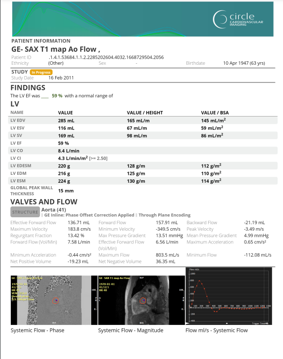
Flow
Produce flow information for the evaluation of systolic and diastolic function. Easily quantify shunts, valve regurgitation and compare multiple vessels.
Tissue
Signal intensity and perfusion analysis involving images acquired with contrast agents are only available for non-clinical use in USA.
Effectively evaluate characteristics of myocardial tissue to inform diagnostic decision making for ischemic and non-ischemic diseases. Quantify enhancement, edema, perfusion defects and iron load from simple acquisition sequences.
cvi42 | Strain
Additional license required.
Quantify myocardial deformation without additional time in the scanner. Increase sensitivity for detection of mild functional abnormalities in contrast to EF alone.
- Quantify global and regional radial, circumferential and longitudinal strain in 2D
- AI-based LV contour detection
- Calculate strain rate, displacement, time to peak strain and displacement, velocity, torsion, and torsion rate
cvi42 | 4D Flow
Additional license required.
Visualizing and quantifying flow patterns anywhere in a 3D structure.
- Preprocessing including offset correction and antialiasing
- Centerline definition for multiple structures
- Various flow visualizations
- Flow assessment in multiple planes Qp:Qs comparison
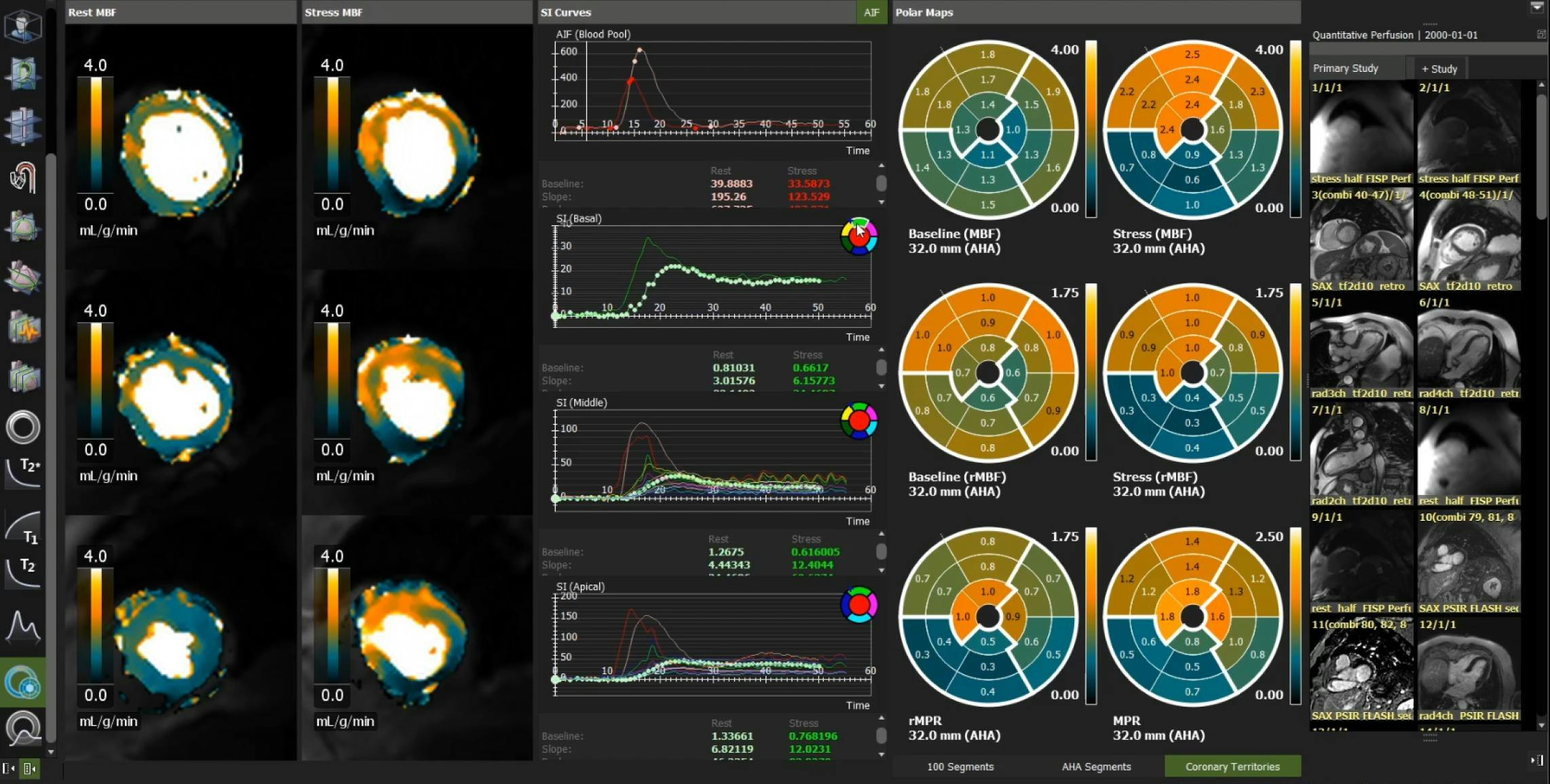
cvi42 | Quantitative Perfusion
Additional license required. Research use only.
Intuitively visualize perfusion defects in patients with suspected Coronary Artery Disease (CAD). Quantify myocardial blood flow at rest and stress to reduce intra-reader variability.
- Streamlined workflow for rest and stress perfusion quantification
- Automated motion correction and contour detection
- Color map display of myocardial blood flow (MBF) values
- Color map display of myocardial perfusion reserve (MPR) values
- Vendor neutral with multi-sequence support
Reporting
cvi42 offers customizable structured reporting for cardiac MR evaluation. Build the report you need for research and daily clinical practice.
Web Viewer
View study images and edit patient reports from any web browser! Access the information you need for an efficient cardiac imaging workflow that works for you.
- Smart CMR viewing with series classification and automated view settings to optimize cardiac imaging
- Apply study tags to organize your cases
- Edit and finalize reports

Why Circle?
Our automated platform increases productivity while streamlining reporting. Reclaim lost time by optimizing decision-making with our cutting-edge AI platform and our unique expertise.

Our Technology Enables Informed Decisions for Fast and Accurate Diagnoses

Automated Workflows Increase Time for Patient Care

Unmatched User Experience & Support You Can Count On

Take the First Step Towards Better Patient Care
Discover today how our tailored solutions can enhance efficiency and productivity while reducing your workload and giving you more time to focus on your patients.